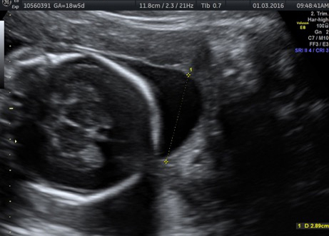
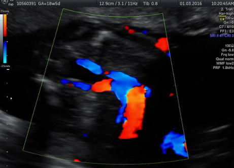
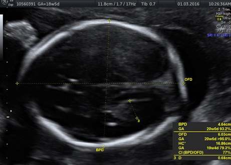
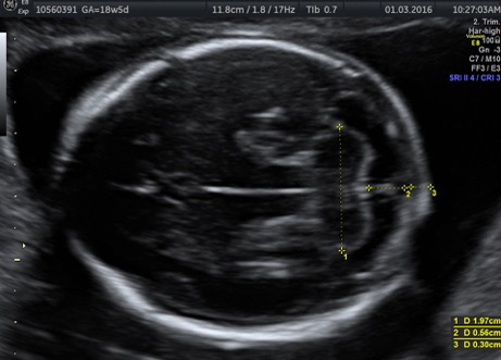
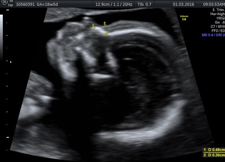
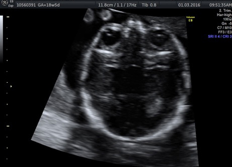
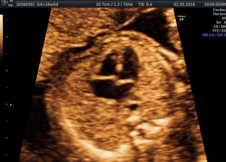
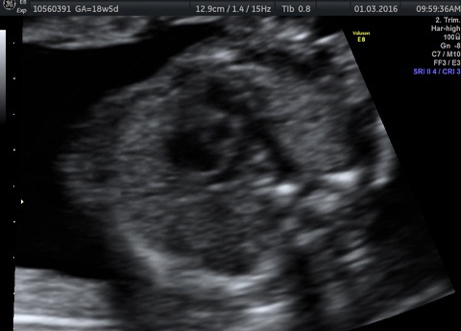
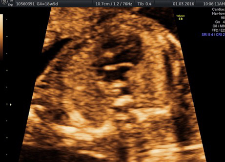
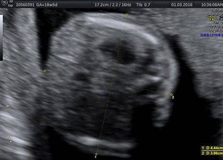
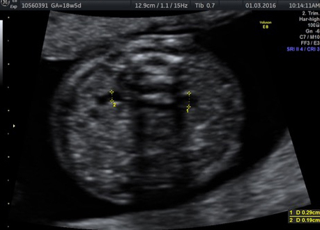
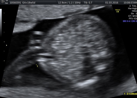
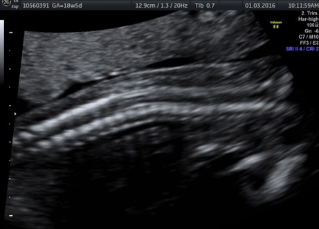
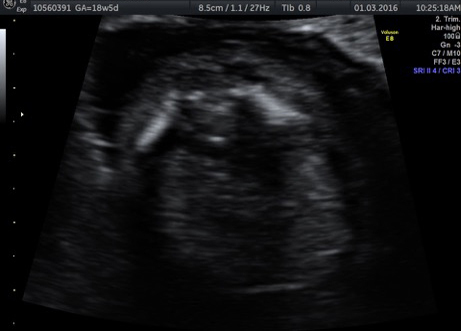
Fetoscopic laser photocoagulation is the preferred method of treatment for certain severe cases of twin-twin transfusion syndrome. The procedure stops the transfer of blood between fetuses, often halting the progression of TTTS. Thorough evaluation, which include including ultrasound and fetal echocardiogram will be conducted and the TTTS staged according to Quintero staging to decide if surgery will be appropriate before deciding if fetoscopic laser photocoagulation is an appropriate option. Generally, the fetus should be between 16 and 26 weeks gestation with no other significant anomalies, and the mother should be healthy and have normal cervical length.
| Quintero Classification for Twin twin transfusion syndrome26 | ||
|---|---|---|
| Stage I | Discordant amniotic fluid in the two sacs of structurally normal fetuses(oligohydramnios defined as DVP < 2 cm; polyhydramnios defined as DVP > 8cm or >10cm at < 20 or > 20 week respectively); Donor bladder visible | |
| Stage II | Donor bladder not visible | |
| Stage III | abnormalities in umbilical artery or ductus venosus in either twin | |
| Stage IV | Fetal hydrops in either twin | |
| Stage V | Intrauterine demise of either twin | |
This is an inpatient procedure. Pre-admission various blood tests will be run to check for infection.
On the day of procedure
Antibiotics and medicines to stop premature uterine contractions will be given before you are brought into the operation theatre.
In the operation theatre there will be the operating team, the OT staff and the anaesthiatist as well as the laser machine, endoscopy stack and the ultrasound machine.
The anaesthesia usually given is local and regional where you will be aware of the procedure but comfortable and not in pain. This is because during the procedure we interact with the mother and may need to give instructions to her to turn in certain directions. Our surgeon makes a small incision in the mother’s abdomen, and a trocar (tiny metal tube) is inserted into the uterus. The fetoscope is passed through the trocar to examine and map the blood vessel connections on the surface of the placenta shared by the twins. The image is viewed on a large computer monitor. Ultrasound is used to continuously monitor the procedure. A laser is used to seal abnormal blood vessels and disconnect them permanently, and the surgeon drains excess amniotic fluid through the trocar before removing it. After sealing the blood vessels between the twins, our surgeons laser a line between the connections in order to coagulate even smaller vessels from one side to the other. This process is called solomonization.
Although each case is different, most patients generally remain in the hospital for one day.
One week after surgery, ultrasound and fetal echocardiography are performed to evaluate the condition of the fetuses.
Although every procedure has risk, the chance of serious side effects with this procedure are minimal. The most common problem is premature labor. During your evaluation, your doctors at the Apollo Centre for Fetal Medicine will provide a detailed review of your baby’s condition and treatment options.


