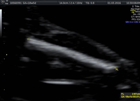
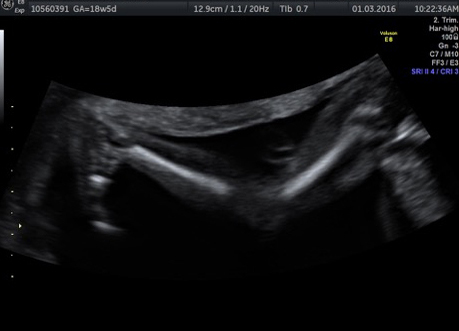
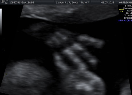
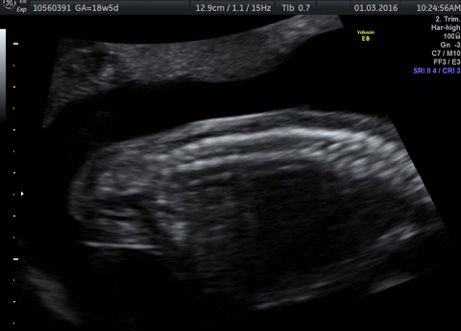
A dedicated fetal MRI service was started in 2008..
Led by Dr. A.S.Arora we have a 3T Philips scanner and take single shot ultra fast sequences generally without sedating the fetus. High resolution imaging within the established safety parameters is the future of fetal diagnostics and Dr. Arora believes that our protocols match the global standards of care..
We are constantly updating our imaging protocols and exploring new avenues which allow 3D reconstruction and better anatomical delineation..
Fetal MRI is the MRI evaluation of the Fetus during pregnancy for specific clinical indications, without causing any risk to fetus and to the ongoing pregnancy.
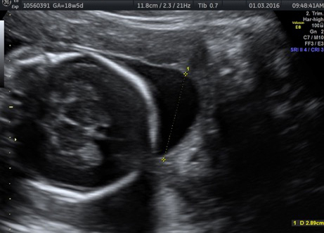
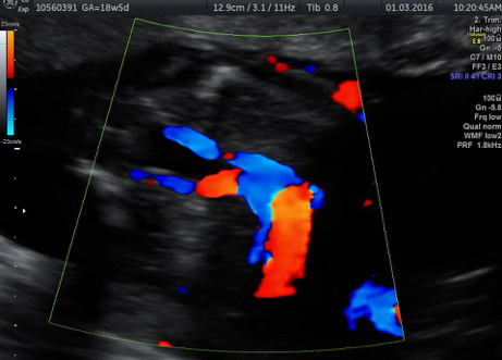
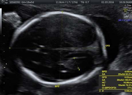
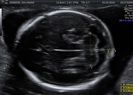
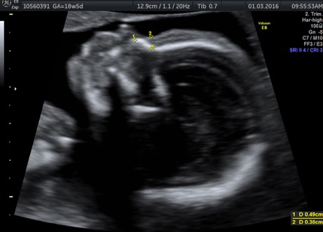
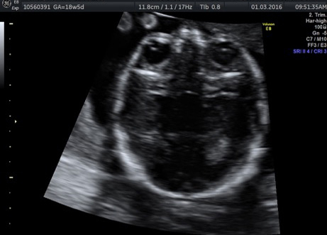
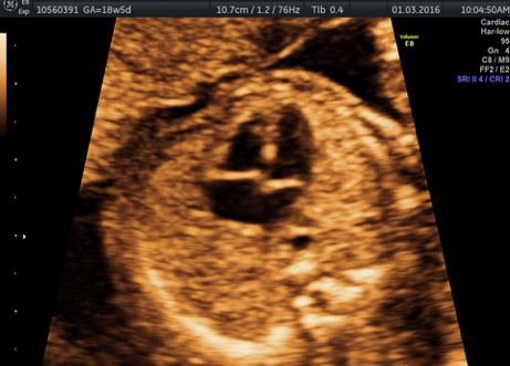
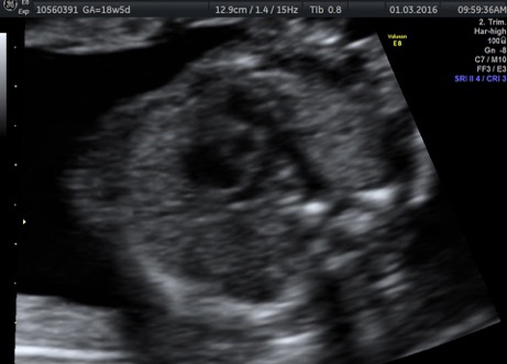
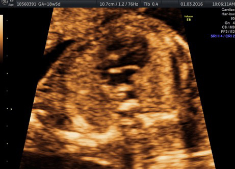
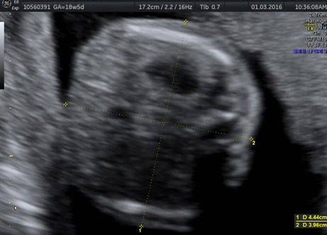
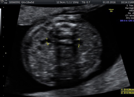
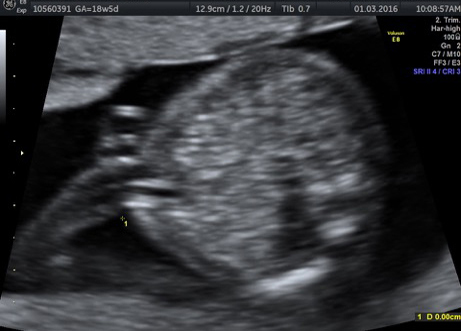
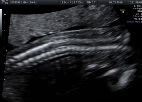
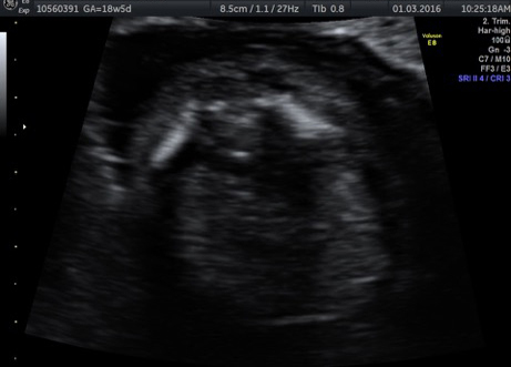
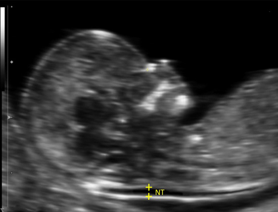
- Ultrasound is certainly the initial imaging modality to assess fetal well-being. There are however, certain circumstances when the visualization of fetus is not optimal. Fetal MRI provides the desired information about the fetal structures in these cases.
- If findings in fetal Ultrasound scan are suspicious or abnormal, MRI is a useful option for further evaluation of fetus. MRI findings may help in confirmation of the abnormality or may provide finer details about the fetal structural anomaly.
- MRI is a particularly useful modality for finer evaluation of the fetal brain. MRI is useful to detect additional lesions which may help in deciding the prognosis and facilitate decision-making (continuation or termination of pregnancy).
- Fetal MRI is usually advised following a level-2 ultrasound (anomaly scan), at around 20-22 weeks. At times, the scan may be deferred till 26-28 weeks, depending upon the clinical indication.
- Fetal MRI may also be advised in the third trimester, following abnormal ultrasound findings or as a follow-up scan.
The mother needs to lay supine or in left lateral position on the MR scan table which then moves inside the MRI machine. The machine and procedure is same as any other MRI scan.
- Fetal MRI should always be properly scheduled. It is better to ask for an afternoon appointment, as fetal movements are usually less during this time.
- There is no need for the Mother to remain fasting for the scan. The mother should have her lunch at least 1-2 hours prior to the time of appointment.
- No injections or oral medicines will be given during or prior to scan.
- Mother need not hold urine for the scan. It is advisable for the mother to void 15-20 min prior to the scan.
- Mobile phones, coins, keys, belts, wrist-watches, jewellery or any form of metal should be left outside the scanning room.
- It is advisable to walk slowly for 10-15 min prior to the MRI scan.
- In accordance with the PC-PNDT act, Fetal MRI will not be used to determine the sex of the baby in any way. The mothers are therefore expected to fill up the relevant forms and duly sign them, prior to the scan.
- Few people may experience extreme claustrophobia due to the closed structure of the MR machine and may not cooperate for the scan. Mild to moderate degree of claustrophobia usually does not cause any problem. It is important to understand that MRI systems are absolutely safe and the scans are always monitored. A relative is also asked to sit in the scanning room, close to the patient for further reassurance.
- As with any MRI examination, the mother will hear a variety of sounds from the MRI machine, during the image acquisition. In case the patient experience any area of localized heating or any area of skin sensation or any kind of discomfort, the patient can raise an alarm with the help of hand switch.
Getting an MRI done in the 2nd and 3rd trimesters of pregnancy is absolutely safe and is approved by all apex medical societies.
- MRI evaluation of fetus provides detailed information about the fetal structures with high accuracy. Complete assessment of fetal heart is however, not feasible with fetal MRI, for which echocardiography is the most accurate modality.
- The protocol for Fetal MRI is often tailored according to clinical indication, which is studied in detail. The rest of the structures are also simultaneously screened. Despite high accuracy, performance of Fetal MRI does depend on the extent of structural defect, fetal movements and the image quality.
- Apart from the fetal structures, the fetal environment including the placenta, amniotic fluid and the umbilical cord can be well evaluated with fetal MRI. Fetal MRI however, cannot diagnose functional disorders of the fetus.
Mothers who had history of cardiac valve placement or any metallic device for fractures cannot undergo fetal MRI. It is therefore important for the patient to inform the concerned Fetal Medicine Specialist, Radiologist & MRI technician about any form of prior surgical procedure.
The appointment for Fetal MRI can be scheduled at the CT-MRI reception at the following phone number: 011-29873018
All the Fetal MRI scans are duly monitored by the Radiologist. Soon after the scan is done, your Radiologist will talk to you and convey the provisional reports. The final detailed report does take time and may be ready either the same day or the next working day. You are free to discuss the report with your Radiologist and the Fetal Medicine Specialist.


